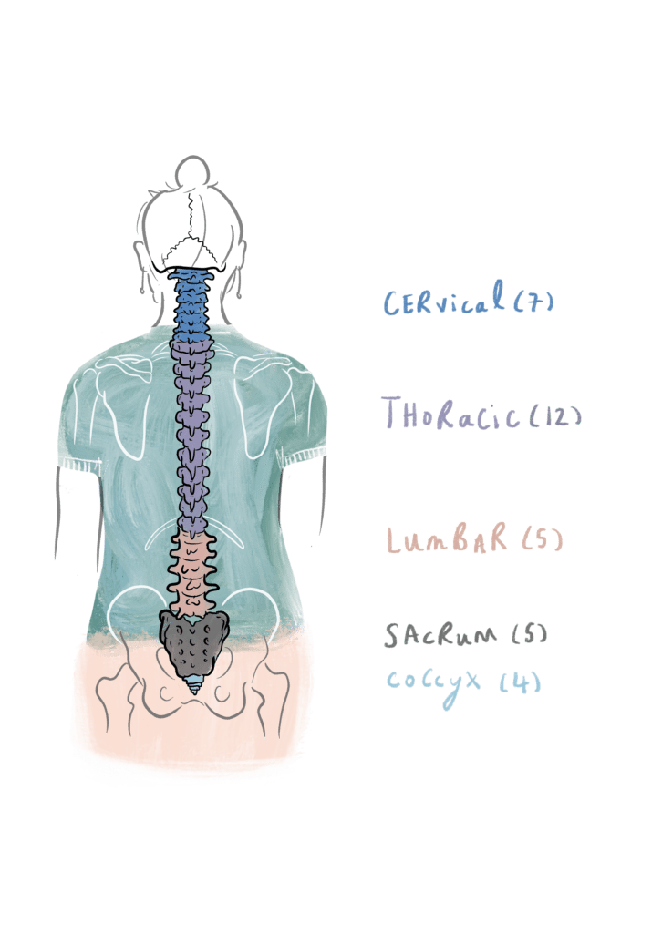
The spine is normally made up of a total of 29 backbones or vertebrae (‘vertebra’ being the term for a single backbone). There are four regions in the spinal column: the neck (made up of seven cervical vertebrae), the chest (made up of twelve thoracic vertebrae), the lower back (made up of five lumbar vertebrae) and the part attached to the pelvis (made up of five sacral vertebrae fused together to form the sacrum).

The backbones in each region of the spine are usually referred to by number going from top to bottom, e.g. the five lumbar vertebrae in this region (going from top to bottom) are L1, L2, L3, L4 and L5 (‘L’ being short for ‘lumbar’). The rest of the vertebrae are named in a similar way (C1, C2, etc for the cervical spine; T1, T2, etc for the thoracic spine; and S1, S2, etc for the sacral spine). Note that the spinal cord usually finishes at the level of approximately L1, meaning that most of the lumbar spine does not actually have spinal cord travelling through it, but just nerves called the cauda equina.

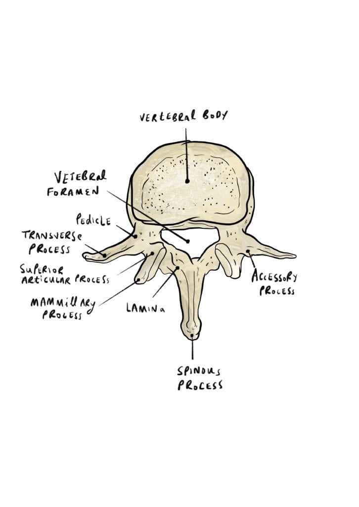
Figure 2 shows one of the lumbar backbones as if you were looking down at it from above. You will notice that each vertebra of the spinal column has a large hole in the middle called the spinal canal. It is through this canal that the spinal cord and nerves pass downwards from the brain to different parts of the body. The spinal canal is surrounded by the vertebral body in front (carrying the weight), the pedicles on the left and right and the lamina behind. In addition, a pair of transverse processes stick out on either side, and there is a single large spinous process protruding backwards from the lamina of each vertebra (which you may be able to feel on your own back as a series of hard knobbles about an inch apart).
The joints between neighbouring backbones are formed by articular facets located on both the top and bottom of each vertebra. In addition to the bony structures of the vertebrae, a number of ligaments are present in the spinal canal, holding the vertebrae together.
Lastly, sandwiched between each vertebral body is an intervertebral disc. Since the function of these discs is to cushion impact between the vertebrae, they are normally soft. The discs normally become more fibrous with age.
Credit

Axum Gebreyohanes
Medical student at University College London
Illustrations

Merlin Strangeway
Medical Illustrator
Merlin is an award-winning Medical Illustrator, Writer, Educator and Director of ‘Drawn to Medicine’. She strives to visualise information in a way that makes it universally accessible, educational and engaging for both clinicians, patients and the general public.

Leave a Reply